
Each year, April is recognized as Oral Cancer Awareness Month, a time dedicated to raising awareness about oral cancer, promoting early detection, and encouraging regular dental checkups. This month, KOHC member Dr. Jennie Ison provides insight into a new trend in oral cancer presentation and tips on how to spot it in your practice.
As an oral pathologist, my primary role is the diagnosis of biopsy specimens taken from both the hard and soft tissues of the oral cavity. While not all of these are concerning for cancer, in many situations, the goal of the biopsy is to do just that: prove that a lump or bump in a patient’s mouth is not, in fact, cancer.
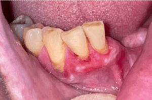
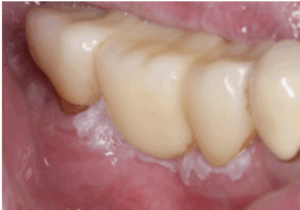
In addition to this, I have the great privilege of educating (and learning from) the future dentists of Kentucky. For years we have been teaching that the most concerning locations for development of cancer in the mouth are the lateral and ventral (sides and bottom) tongue and the floor of the mouth (the area under the tongue). While this certainly remains true, anecdotally, I have noticed an increase in oral cancer occurring in another, sneakier location, one that may go undiagnosed for an extended period of time: the gingiva. At the recent annual meeting of the American Academy of Oral and Maxillofacial Pathology, discussions with my colleagues from around the United States and other countries yielded the same observation.
Why is this? We still need more concrete data on the who and why, but one of the reasons it can fly under the radar, ultimately leading to a delay in diagnosis and treatment, is that it likes to mimic benign, reactive, or inflammatory lesions, especially periodontal disease, pericoronitis, and papillary epithelial lesions, such as squamous papillomata. You’ve probably heard “If you hear hoofbeats, think horses, not zebras”, indicating that common things are common. However, when a lesion doesn’t respond to reasonable therapy, have a low threshold to recommend or seek biopsy.
Features to be on the look-out for:
- Single tooth mobility/bone loss
- A well-demarcated white lesion that doesn’t wipe away
- Any localized red area that can’t be attributed to inflammation or doesn’t respond to a single round of appropriate antibiotics or antifungal medications
- Any rapidly growing area
- An ulcer that doesn’t heal within 2 weeks
Join KOHC as we continue to spread awareness and provide resources – like this fact sheet for at-home screening – during Oral Cancer Awareness Month and throughout the year.


